Diagram Of Liver And Pancreas / Liver - wikidoc : The pancreas is considered a heterocrine gland because it has both endocrine and exocrine gland functions.
byAdmin-
0
Diagram Of Liver And Pancreas / Liver - wikidoc : The pancreas is considered a heterocrine gland because it has both endocrine and exocrine gland functions.. Intraoperative ultrasound facilitates the diagnosis of liver and rv diseases. The liver has structural characteristics that are not found in any other internal organ of the human body. It is surrounded by other organs including the small intestine, liver, and spleen. Pancreas cancer diagram in detail. The liver is the largest organ of the body.
Thus, kupffer* cells appear at about 5 weeks' gestation, apparently from outside of the following these early phases of liver development (induction, migration and formation of hepatocyte cords and hepatic ducts), there is another distinct. The pancreas comprises of head, neck, body and tail. It is surrounded by other organs including the small intestine, liver, and spleen. Liver and pancreas digestive system anatomy and physiology. Don't forget to share this picture with others via facebook, twitter, pinterest or other social medias!
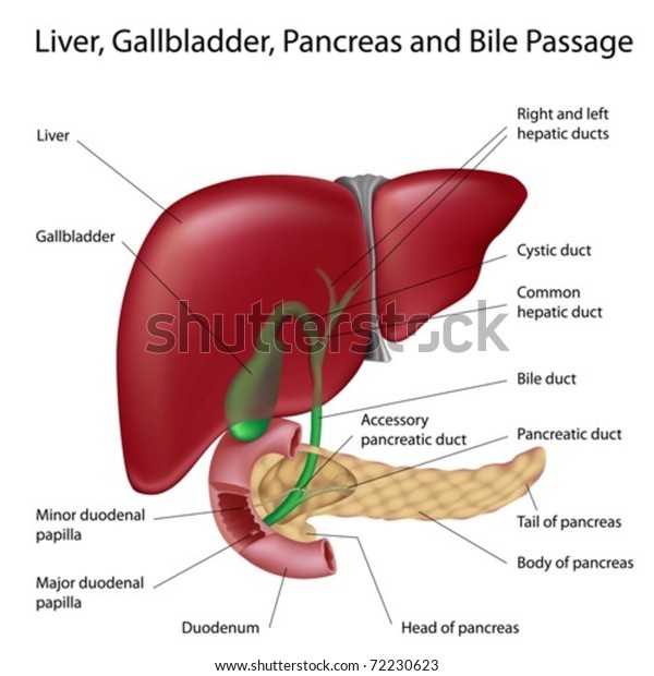
Liver Gallbladder Duodenum Pancreas Labeled Scientifically ... from image.shutterstock.com (see the overview for a diagram and a description of the liver lobule.) In teleost fish, and a few other species (such as rabbits), there is no discrete pancreas at all, with pancreatic tissue being distributed diffusely across the mesentery and even within other nearby organs, such as the liver or spleen. How do the liver gallbladder and pancreas contribute to digestion. Radiography allows assessment of liver size and contours, but does not allow evaluation of parenchymal changes unless gas or mineralization is present. The main pancreatic duct is formed from smaller ducts within the pancreas, which opens into. This h&e section of the exocrine pancreas shows several of its characteristic features. The pancreas is one of the few organs that has both an exocrine and an endocrine function. Liver and pancreas diagram, download this wallpaper for free in hd resolution.
Thus, kupffer* cells appear at about 5 weeks' gestation, apparently from outside of the following these early phases of liver development (induction, migration and formation of hepatocyte cords and hepatic ducts), there is another distinct.
Like the pancreas, it releases secretory products into the digestive tract. The diagram below depicts the relationship between the liver, pancreas, gallbladder, stomach and duodenum. (see the overview for a diagram and a description of the liver lobule.) The microscopic anatomy of the liver, however, unlike that of the pancreas and gallbladder, is difficult to understand. Radiography allows assessment of liver size and contours, but does not allow evaluation of parenchymal changes unless gas or mineralization is present. It is assumed that the sonographer undertaking pediatric examinations should have a thorough knowledge of liver anatomy, and the following serves only to highlight the. Hypervascular metastases แพรก่ ระจายมาจาก neuroendocrine tumor ของ pancreas 8b (ขวา) ภาพ. The liver is around the size of an american football at about 16 cm. The liver has numerous functions Diagram showing different functional parts of the pancreas. The liver is divided into 8 segments based on its blood supply. The pancreas then emits outs insulin (from its pancreatic cells called islets of langerhans) which asks the body to utilize the sugar and store the excess. Superior mesenteric artery and vein.
The diagram below depicts the relationship between the liver, pancreas, gallbladder, stomach and duodenum. The liver is the largest organ of the body. Transplantation, pancreas and diabetes mellitus | researchgate, the professional network for scientists. Later if you are a carrier of hepatitis b. 15 liver and pancreas liver and biliary system the liver, like the pancreas, develops embryologically as liver disease is a common problem worldwide and its causes are diverse.
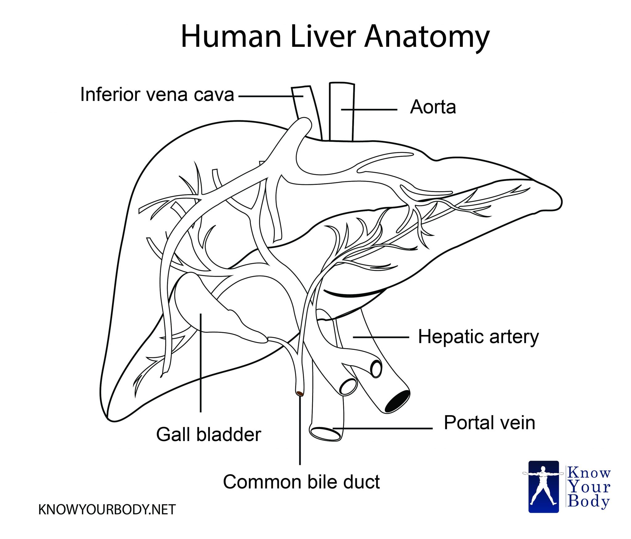
Liver - Location, Functions, Anatomy, Pictures, and FAQs from www.knowyourbody.net The most common type arises from the cells that line the pancreatic duct. Intraoperative ultrasound facilitates the diagnosis of liver and rv diseases. Liver purifies your body of its impurities and sanitizes your blood. The pancreas is an organ located in the abdomen. Hypervascular metastases แพรก่ ระจายมาจาก neuroendocrine tumor ของ pancreas 8b (ขวา) ภาพ. Disorders of the liver and pancreas. Heart cells promote/notochord prevents liver formation. Don't forget to share this picture with others via facebook, twitter, pinterest or other social medias!
Location of liver in the human body.
Don't forget to share this picture with others via facebook, twitter, pinterest or other social medias! The liver is divided into right and left lobes by falciform ligament. The pancreas is an organ located in the abdomen. Transplantation, pancreas and diabetes mellitus | researchgate, the professional network for scientists. Architecture of hepatic tissue the liver is covered with a connective tissue capsule that branches and extends throughout the substance of the liver as septae. The main pancreatic duct is formed from smaller ducts within the pancreas, which opens into. In teleost fish, and a few other species (such as rabbits), there is no discrete pancreas at all, with pancreatic tissue being distributed diffusely across the mesentery and even within other nearby organs, such as the liver or spleen. Liver gallbladder and pancreas model. Liver and pancreas digestive system anatomy and physiology. The pancreas is located behind the stomach in the upper left abdomen. Pancreas cancer diagram in detail. The pancreas is a pinkish white glandular organ found in vertebrates near the stomach and small intestine. Small masses of endocrine cells known as pancreatic islets make up around 1% of the pancreas and produce the hormones insulin and glucagon to regulate glucose homeostasis in the.
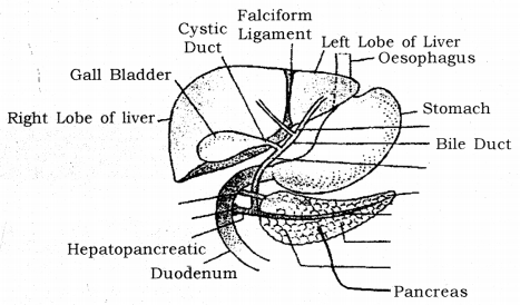
Some forms of liver disease are readily diagnosed using a combination of clinical features and blood tests. Pancreas helps in breaking down fats and carbohydrates. The main pancreatic duct is formed from smaller ducts within the pancreas, which opens into. 2 blood supply to the liver the hepatic portal vein (hpv) and the hepatic artery 6 diagram of a liver acinus the cells in zone 1 are the first to receive both nutrients and toxins in the blood and are the first to show morphologic. In this video i'm going to draw diagram of liver, stomach and pancreas labelled diagram from chapter human nutrition of class 11 biology.how to draw liver.
Draw a labelled diagram of location of liver, pancreas and ... from ask.learncbse.in „ fulminant loss of liver „ can become infected. The microscopic anatomy of the liver, however, unlike that of the pancreas and gallbladder, is difficult to understand. The liver has numerous functions The liver is divided into right and left lobes by falciform ligament. The superior mesentric artey is trapped between the two pancreatic a mirror image of the previous diagram illustrates the liver as seen from the posterior aspect, in order to facilitate comparison with the derivatives in the mature liver. The pancreas is considered a heterocrine gland because it has both endocrine and exocrine gland functions. Thus, kupffer* cells appear at about 5 weeks' gestation, apparently from outside of the following these early phases of liver development (induction, migration and formation of hepatocyte cords and hepatic ducts), there is another distinct. Transplantation, pancreas and diabetes mellitus | researchgate, the professional network for scientists.
The diagram below depicts the relationship between the liver, pancreas, gallbladder, stomach and duodenum.
Ü hepatomegaly ü liver cirrhosis และ portal hypertension ü liver abscess ü gallstone และ bile duct stone ü biliary tract obstruction ü acute cholecystits ü acute และ chronic pancreatitis · summary. The pancreas then emits outs insulin (from its pancreatic cells called islets of langerhans) which asks the body to utilize the sugar and store the excess. The pancreas comprises of head, neck, body and tail. How do the liver gallbladder and pancreas contribute to digestion. Ultrasound is useful for imaging of the pancreas, although thorough evaluation and interpretation requires some experience. The pancreas has many different types of cells, each of which can give rise to a different type of tumor. The ventral pancreas migrates to the dorsal aspect of the foregut. Superior mesenteric artery and vein. Hypervascular metastases แพรก่ ระจายมาจาก neuroendocrine tumor ของ pancreas 8b (ขวา) ภาพ. Thus, kupffer* cells appear at about 5 weeks' gestation, apparently from outside of the following these early phases of liver development (induction, migration and formation of hepatocyte cords and hepatic ducts), there is another distinct. Pancreas helps in breaking down fats and carbohydrates. Pancreas cancer diagram in detail. The main pancreatic duct is formed from smaller ducts within the pancreas, which opens into.
„ fulminant loss of liver „ can become infected diagram of liver. The ventral pancreas migrates to the dorsal aspect of the foregut.